


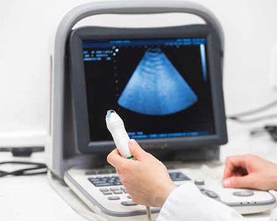
Lung ultrasound as a diagnostic method of lung diseases has been rapidly developing for several decades. It is the first imaging study with such a wide range of mobility. Ultrasound diagnosis of lung diseases of patients of all ages is increasingly used in medical practice. In order to systematise knowledge in this field, it was necessary to create recommendations on lung ultrasound.
Lung ultrasonography is a diagnostic tool known for several decades. It owes its popularity to the work of many clinicians who use this method of lung imaging on a daily basis in their medical practice with diagnostic successes. Despite the long history of the use of ultrasound in medicine, the lungs did not constitutean attractive medium for propagating the ultrasound wave. The air in the lungs causes the ultrasound wave to be strongly reflected and prevents ‘looking’ into the lung. The result of this valuable observation was the general view that ‘ultrasound is not suitable for lung assessment.’ At the same time, the diagnostics of ‘airless’ parenchymal organs was intensively developed. Hence, ultrasound of the abdominal organs, thyroid gland, nipple, lymph nodes, and the heart are highly specialised.
Initial skepticism towards lung ultrasound was justified and was related to the inability to assess the parenchyma of the air lung properly. It was not until the 1990s that reports appeared on the diversity of the pulmonary changes observed in ultrasound and on the possibility of differentiating the causes of dyspnea. It turned out that with the loss of lung aeration, two types of changes appear in ultrasound: artefacts and consolidations. Both groups of pulmonary changes indicate the degree of lung aeration loss. A careful assessment of changes in the surface of the lung (the so-called pleural line) and subpleural consolidations (areas of the airless lung) allow for the differentiation of the causes of dyspnea. With the passage of time and the work of numerous other scientists, the high accuracy of lung ultrasound in the diagnosis of such diseases as: pneumonia, cardiogenic and non-cardiogenic pulmonary edema, atelectasis, pulmonary infarction changes in the course of pulmonary embolism, pulmonary fibrosis, or pneumothorax and fluid in the pleural cavities has been proven.
The databases of medical literature currently collect several thousand publications on the use of lung ultrasound in anaesthesiology, intensive care, cardiology, pulmonology, paediatrics, and emergencies. With the increasing number of papers, it was necessary to organise the knowledge of lung ultrasound. For this purpose, the first recommendations were created by multinational experts in the field of lung ultrasound in 2012. (Volpicelli G et al.). The first recommendations concerned the use of lung ultrasound as a method dedicated to clinicians, and, therefore, used as point of care diagnostics. In line with EBM rules, the first Recommendation Document was produced. The next documents were created by a group of Polish scientists and practitioners dealing with lung ultrasound on a daily basis. A group of adult health specialists created recommendations for internal medicine specialists in 2017, which were updated in August 2020. Recommendations for Lung Ultrasound in Internal Medicine is the title of publications in the journal Diagnostics (Buda N et al.). The document is updated with the latest scientific data. The content contains information on pathological changes in the lungs (i.e. pneumothorax, edema, fibrotic interstitial lung diseases, inflammatory, atelectasis, neoplastic changes, as well as pulmonary embolism). Recommendations regarding cardiogenic pulmonary edema and pulmonary congestion in patients undergoing dialysis have been particularly enriched. The sonomorphology of the above mentioned changes, as well as the accuracy of lung ultrasound in their diagnosis compared to the chest X-ray examination, were thoroughly assessed in terms of the quality of available literature (in the GRADE system) and the opinion of the working group (in the Delphi system). Moreover, the recommendations also refer to the examination protocol, examination technique and education of students and doctors in the field of lung ultrasound. The document presents a complementary approach to the possibilities of lung ultrasound in the technical, clinical and educational context.
At the same time, in Poland, a group of paediatricians was working on a document relating to the use of lung ultrasound in paediatrics. Therefore, in 2020, a consensus was also published on the use of lung ultrasound in paediatric patients. The document, entitled: "Consensus on the Application of Lung Ultrasound in Pneumonia and Bronchiolitis in Children" (Jaworska J et al.), addresses the possibilities, validity and terminology of lung ultrasound in children with lower respiratory tract infections. Both documents present the possibilities of using a non-invasive examination such as lung ultrasound in the diagnosis of both paediatric and adult patients.
Since March 2020, the use of lung ultrasound has gained publicity again, due to its use as a diagnostic tool in the fight against the COVID-19 pandemic. Doctors and paramedics, who are in the first line of contact with the patient, began to have portable, compact ultrasound machines. Pocked size ultrasound scanners can be used at any place of the incident: at the patient's home, at the site of a traffic accident, during patient transport, as well as in emergency departments and specialist departments of a hospital. Numerous educational materials from around the world appeared on the web: publications, on-line courses and lectures aimed at educating medical personnel and preparing them to work independently with an ultrasound machine during the COVID-
19 pandemic. However, it should be noted that lung ultrasound, as well as chest tomography, does not detect the SARS-COV-2 virus, which causes the COVID-19 disease. As a diagnostic tool, lung ultrasound can show interstitial inflammatory lesions, e.g. in the course of COVID-19, influenza, and atypical infections. In order to confirm the cause of interstitial inflammatory lesions in the lungs visible in the imaging test, we use PCR testing for SARS COV-2 infection or antigen tests. Access to chest tomography is difficult in many places where lung inflammatory changes in the course of COVID-19 are diagnosed. In such a situation there are: intensive care units, some COVID-19 dedicated wards, but also all pre-hospital care, including an ambulance service. Due to the needs of clinicians, another document for the needs of intensive care was created, entitled "Consensus of the Study Group for Point-of-Care Lung Ultrasound in the intensive care management of COVID-19 patients" (Buda N et al.) Published in Anaesthesiology Intensive Therapy, 2020.
As evidence shows, lung ultrasound is becoming the ‘stethoscope of the 21st century’. It is the first imaging study with such a wide range of mobility. Ultrasound diagnosis of lung diseases of patients of all ages is used more and more often and more readily accepted by the scientific community. In 2019, ERS issued a document entitled: "ERS statement on chest imaging in acute respiratory failure" (Chiumello et al.). The ERS clearly presents its position on lung ultrasound in the diagnosis of acute respiratory failure. According to the ERS expert group, lung ultrasound is a method that is more effective than chest X-ray and is comparable to chest tomography, after proper training of the person performing the ultrasound examination.
Literature:
Volpicelli, G.; Elbarbary, M.; Blaivas, M.; Lichtenstein, D.A.; Mathis, G.; Kirkpatrick, A.W.; Melniker, L.; Gargani, L.; Noble, V.E.; Via, G., et al. International evidencebased recommendations for point-of-care lung ultrasound. Intensive Care Med 2012, 38, 577-591, doi:10.1007/s00134-012-2513-4.
Buda, N.; Kosiak, W.; Radzikowska, E.; Olszewski, R.; Jassem, E.; Grabczak, E.M.; Pomiecko, A.; Piotrkowski, J.; Piskunowicz, M.; Soltysiak, M., et al. Polish recommendations for lung ultrasound in internal medicine (POLLUS-IM). J Ultrason 2018, 18, 198-206, doi:10.15557/JoU.2018.0030.
Buda, N.; Kosiak, W.; Wełnicki, M.; Skoczylas, A.; Olszewski, R.; Piotrkowski, J.; Skoczyński, S.; Radzikowska, E.; Jassem, E.; Grabczak, E.M., et al. Recommendations for Lung Ultrasound in Internal Medicine. Diagnostics (Basel) 2020, 10, doi:10.3390/diagnostics10080597.
Jaworska J ,Komorowska-Piotrowska A, Pomiećko A, WiśniewskiJ, Woźniak M, Littwin B, Kryger M , Kwaśniewicz P, Szczyrski J, Kulińska Szukalska K, Buda N, Doniec Z, Kosiak W; Consensus on the Application of Lung Ultrasound in Pneumonia and Bronchiolitis in Children Diagnostics (Basel) . 2020 Nov 11;10(11):935. doi: 10.3390/diagnostics10110935.
Guan WJ, Ni ZY, Hu Y et al. Clinical characteristics of coronavirus disease 2019 in China. N Engl J Med. 2020. doi:10.1056/NEJMoa200203
Ai T, Yang Z, Hou Het al. Correlation of Chest CT and RT-PCR Testing in Coronavirus Disease 2019 (COVID-19) in China: A Report of 1014 Cases. Radiology. 2020 Feb 26. doi: 10.1148/radiol.2020200642
Buda N, Andruszkiewicz P, Czuczwar M, Gola W, Kosiak W, Nowakowski P, Sporysz K. Consensus of the Study Group for Point-of-Care Lung Ultrasound in the intensive care management of COVID-19 patients.Anaesthesiol Intensive Ther. 2020;52(2):83-90. doi: 10.5114/ait.2020.96560.
Chiumello D, Sferrazza Papa GF, Artigas A, Bouhemad B, Grgic A, Heunks L, Markstaller K, Pellegrino GM, Pisani L, Rigau D, Schultz MJ, Sotgiu G, Spieth P, Zompatori M, Navalesi PERS statement on chest imaging in acute respiratory failure. European Respiratory Journal 2019; DOI: 10.1183/13993003.00435-2019


